TIGEM researchers are studying HITI genome editing to develop stable and durable treatments for genetic diseases affecting damaged liver tissue.
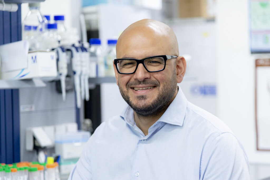
There is a fundamental difference between developing a technology and understanding the conditions under which it can become a viable therapy. Pasquale Piccolo’s research focuses on this crucial second step. A Group Leader at TIGEM and Associate Professor of Medical Genetics at the University of Naples Federico II, Piccolo uses HITI — a genome-editing strategy developed at TIGEM by Alberto Auricchio's laboratory — to investigate its potential application to specific genetic liver diseases.
HITI, or Homology-Independent Targeted Integration, enables the stable insertion of a therapeutic sequence into the genome by harnessing a DNA-repair mechanism that remains active even in non-dividing cells. Piccolo’s group is investigating the disease settings in which these features could provide a meaningful therapeutic advantage.
Because of its central role in metabolism and in the production of circulating proteins, the liver is one of the main target organs for gene therapy. Piccolo’s research, however, focuses on a particular group of conditions: diseases in which the genetic defect does not merely cause a systemic deficiency, but also directly damages hepatocytes.
In these cases, the liver is not simply the organ to which the therapy must be delivered. It is already a tissue profoundly altered by the disease itself.
This creates challenges for gene therapy and genome editing that are very different from those encountered in an otherwise healthy liver. The innovative aspect of Piccolo’s work lies in applying HITI to these specific diseases and developing strategies designed to maintain efficacy, durability and safety even when the target organ is already compromised.
Finding the right therapy when the liver is damaged
The liver is a particularly important target for gene therapy because it produces many proteins that act in the bloodstream or in other organs. In haemophilia, for example, treatment is delivered to hepatocytes, even though the underlying genetic defect does not directly damage the liver.
The diseases studied by Piccolo’s team at TIGEM are different. In these conditions, the mutation disrupts essential functions within liver cells and causes direct damage to the organ itself.
Gene therapy and genome editing therefore face a profoundly different biological environment in a damaged liver than in healthy tissue. Inflammation leads to the activation and accumulation of immune cells; the deposition of collagen and other components of the extracellular matrix causes fibrosis; and the death of hepatocytes triggers a regenerative response, prompting the surviving cells to proliferate.
These changes can directly affect treatment delivery, the immune response and the durability of the therapeutic effect.
Fibrosis alters the architecture of the tissue, making it more difficult for vectors to reach hepatocytes. Immune cells accumulating in the damaged liver may also reduce treatment efficiency by capturing some of the vectors before they reach their target. At the same time, they may increase the likelihood of an immune response against the therapy.
Hepatocyte proliferation creates a different and more specific challenge for conventional gene therapy based on the delivery of a functional copy of a gene through adeno-associated virus, or AAV, vectors. Once released into the nucleus, the vector genome remains largely episomal, meaning that it is separate from the cell’s chromosomes.
When hepatocytes divide, the therapeutic DNA is not replicated together with the cellular genome and is therefore progressively diluted. This can occur both in the growing paediatric liver and in the adult liver undergoing chronic regeneration. As clinical experience has also shown, the result may be a gradual loss of therapeutic benefit over time.
In these diseases, the stability of the genetic correction is therefore not simply desirable: it becomes a core requirement of the therapeutic strategy. HITI is designed to address this limitation by integrating the therapeutic sequence directly into the genome of hepatocytes.
HITI: targeted and stable integration into the genome
HITI is designed to meet two key requirements: making the genetic correction durable over time and directing it towards a precise location in the genome. To achieve this, the platform combines the CRISPR-Cas system with a donor DNA sequence carrying the therapeutic genetic material to be integrated.
Cas is a nuclease, an enzyme capable of cutting DNA. It is guided to the region that needs to be modified by a guide RNA: one part interacts with Cas, while another is complementary to the target DNA sequence. The same nuclease can therefore be redirected towards different genomic loci simply by changing the guide RNA.
Once the DNA has been cut, HITI takes advantage of one of the cell’s natural repair pathways: non-homologous end joining, or NHEJ. Unlike homology-based repair mechanisms, which operate mainly in dividing cells, NHEJ is also active in non-dividing cells. This makes it particularly relevant for tissues such as the liver, where many hepatocytes are normally quiescent.
The donor DNA is also engineered to favour integration in the correct orientation. When it is inserted in the direction required for transgene expression, the Cas recognition sites are not reconstituted, so the region is not cut again. If integration occurs in the opposite orientation, however, the recognition sites are restored. Cas can then cut the sequence once more, creating another opportunity for correct insertion.
This mechanism increases the likelihood of obtaining functional integrations.
“The advantage of a genome-editing platform such as HITI lies in its ability to integrate into the host genome and therefore ensure the persistence of the therapeutic transgene” Piccolo explains.
In the diseases studied by his group, this means that the integrated sequence can be retained even as hepatocytes proliferate. HITI can therefore overcome one of the main limitations of episomal vector genomes, which are progressively diluted as cells divide.
In the liver, the group primarily targets the albumin locus, one of the most highly expressed genes in hepatocytes. Integrating the therapeutic sequence into this region makes it possible to exploit its strong transcriptional activity and produce high levels of the therapeutic protein. In the mouse models studied so far, this approach has not caused any significant reduction in the production of endogenous albumin.
When gene expression requires more precise regulation, however, the donor sequence can instead be directed towards the endogenous locus associated with the disease. This allows the therapeutic gene to remain under the control of its natural regulatory mechanisms.
The ability to select the target locus means that HITI can potentially be applied to both recessive and dominant disorders. In recessive diseases, integration can restore a missing function. In dominant gain-of-function conditions, HITI may instead combine the inactivation of the mutated copy with the introduction of a healthy sequence.
In the most translational configuration currently being investigated by the group, the donor DNA is delivered to the nucleus through an AAV vector, while nanoparticles carry the mRNA encoding Cas. The nuclease is therefore produced only transiently within the cell, reducing the risks associated with prolonged Cas expression.
Hepatocyte proliferation becomes an advantage
The ability to operate in non-dividing cells broadens HITI’s potential applications, but it does not remove one of the main limitations of in vivo genome editing: efficiency. Modifying cells directly inside the body is considerably more complex than working ex vivo, where cells can be isolated, assessed and selected before being returned to the patient.
“One of the main challenges is therefore efficiency, which in vivo — at least in mice — does not exceed 11–12%” Piccolo explains.
This level of editing may be sufficient when a relatively small number of corrected cells can produce large amounts of a therapeutic protein. In many liver diseases, however, a higher proportion of corrected cells is needed to achieve a meaningful benefit.
In the conditions studied by Piccolo’s group, this limitation may be partly overcome by a feature of the diseases themselves: corrected hepatocytes gain a proliferative advantage over diseased cells.
The genetic defect damages hepatocytes and makes them more likely to die. By contrast, cells in which HITI restores the missing function survive for longer and contribute to liver regeneration. Over time, while uncorrected hepatocytes continue to be lost, corrected cells expand and progressively repopulate the tissue.
An initial correction rate of around 10% may therefore increase until it reaches a therapeutically effective threshold.
The group has observed this process in two recessive liver diseases. The first is progressive familial intrahepatic cholestasis type 3, or PFIC3, a condition primarily associated with impaired bile acid metabolism. In this model, HITI has already produced proof of efficacy in animals: the initial level of correction, although insufficient on its own, is progressively amplified by the expansion of corrected hepatocytes.
The second condition is Wilson disease, which is caused by an alteration in copper metabolism. Here too, hepatocytes in which the genetic function has been restored acquire an advantage over diseased cells and expand over time.
In both models, the initial editing efficiency is not sufficient by itself to correct the disease. It is the subsequent expansion of corrected cells that can turn a partial genetic modification into a potential therapeutic benefit.
Hepatocyte proliferation, which is normally a threat to the durability of gene therapy, thus becomes a mechanism for amplifying its effect.
The challenges of genome editing for liver diseases
Stable integration addresses one of the problems created by hepatocyte proliferation: it prevents the therapeutic sequence from being progressively diluted as the liver regenerates. It does not, however, remove the other obstacles associated with liver damage.
Many patients are diagnosed only after inflammation, fibrosis or more advanced tissue alterations have already developed. These conditions may affect both the efficiency and the safety of treatment.
One of the first challenges is delivering the donor DNA to hepatocytes. Piccolo’s group has found that fibrosis can significantly reduce the efficiency of some AAV vectors, including vectors already used in clinical settings. The extent of this loss depends on the capsid, the protein shell that surrounds the vector.
TIGEM is therefore working within a European consortium to engineer and select capsids that are better suited to operating in damaged tissue. This research stems from the observation that some capsid variants are strongly affected by changes in liver architecture, while others retain a good capacity to transfer genetic material even in a fibrotic liver.
Alongside efficiency, immunological safety remains an open question. Current knowledge of AAV vectors is based on their use in thousands of patients, but mainly in people without significant liver damage.
“The vast majority, if not almost all of them, did not have a compromised liver” Piccolo notes.
Fibrosis and liver disease are, in fact, exclusion criteria in most clinical trials. It is therefore still unclear whether inflammation and the presence of activated immune cells alter the response to the vectors or increase specific risks.
The group is investigating how liver damage may change the immunological profile of AAV-based treatments and whether evidence obtained in healthy livers can be fully applied to patients whose target organ is already compromised.
A further layer of complexity concerns genomic safety. HITI is designed to direct the donor DNA towards a defined genomic locus.
“In an ideal setting, the vector would integrate exclusively at the intended site, but we know that this is not what happens in practice” Piccolo explains.
Off-target integration events may occur, as they can with all genome-editing platforms. In a damaged liver, however, these events may differ from those observed in healthy tissue.
Chronic injury alters the transcriptional profile of hepatocytes: some genes are activated, others change their level of expression, and different regions of the genome become more or less accessible. As a result, the distribution of the sites at which vector DNA tends to integrate may also change.
In diseases where corrected hepatocytes have a proliferative advantage, even an off-target integration may be selected over time. If the integration allows the therapeutic sequence to be expressed, the cell may survive and expand within the tissue.
Outside the intended locus, however, the transgene may be expressed in fusion with other sequences, potentially generating a non-canonical protein. The group is therefore assessing how much these events contribute to the therapeutic effect and what risks they may entail.
The implications of this work extend beyond HITI. The condition of the liver can influence the efficiency and safety of all liver-directed gene therapies, including AAV-based treatments that are already available.
For this reason, pre-treatment assessment is no longer limited to liver enzymes and other blood-based markers. Many protocols now include specific tests for fibrosis, such as elastography and, when necessary, biopsy. Establishing whether fibrosis is present, and how advanced it is, can help determine whether a patient is eligible for treatment and allow a more accurate assessment of the balance between potential benefits and risks.
Despite these challenges, the group has already gathered encouraging evidence on both fronts. Vectors equipped with suitable capsids can reach hepatocytes even in a fibrotic liver, while in models of PFIC3 and Wilson disease, the proliferative advantage of corrected cells has amplified the effect of an initially partial level of editing over time.
The innovative potential of the strategy lies in this combination: a targeted and durable correction delivered to an already compromised organ, whose regenerative response may then amplify the therapeutic effect.
The research is now at a preclinical stage of translational optimisation. The group is selecting capsids that perform more efficiently in fibrotic liver tissue, refining the hybrid system that combines AAV vectors with nanoparticles, and characterising the associated immunological and genomic risks.
“We have shown that, with the appropriate adjustments and the right delivery tools, effective gene transfer can be achieved even in the presence of liver damage” Piccolo says.
The ultimate aim is to develop a therapy that can preserve efficacy, durability and safety under the real clinical conditions in which patients are treated.
